1. Introduction
Craniopharyngiomas are rare, benign intracranial tumors that originate from the remnants of Rathke’s pouch, an embryonic structure involved in the development of the pituitary gland. These tumors are situated in the sellar and suprasellar region, near vital structures such as the hypothalamus, pituitary gland, optic nerves, and the circle of Willis. Craniopharyngiomas can be classified into two histological subtypes: adamantinomatous and papillary. The adamantinomatous subtype is more common in children, while the papillary subtype occurs predominantly in adults.
Though these tumors are benign and grow slowly, their close proximity to critical brain structures can lead to significant neurological, hormonal, and visual impairments. Craniopharyngiomas frequently have both solid and cystic components, which can make their complete surgical resection challenging. These tumors may recur even after surgical removal, necessitating long-term monitoring and follow-up care.
2. Causes
The exact cause of craniopharyngiomas remains unclear, as no specific genetic or environmental factors have been conclusively linked to their development. However, researchers believe that these tumors arise from the embryonic remnants of Rathke’s pouch or the craniopharyngeal duct, structures that play a crucial role in the formation of the pituitary gland during early embryonic development.
Rathke’s pouch is an invagination of the oral ectoderm that gives rise to the anterior lobe of the pituitary gland, while the craniopharyngeal duct is a transient channel connecting Rathke’s pouch to the oral cavity. In some cases, these embryonic remnants may not regress as they should during development, leading to the formation of craniopharyngiomas later in life. Despite this connection, the exact cellular and molecular mechanisms driving the development and growth of craniopharyngiomas are not well understood.
Recent studies have identified some genetic mutations and molecular pathways that may play a role in the development of craniopharyngiomas. For instance, mutations in the CTNNB1 gene have been found in a majority of adamantinomatous craniopharyngiomas, while alterations in the BRAF gene have been observed in a significant proportion of papillary craniopharyngiomas. However, further research is needed to fully understand the role of these genetic changes and how they contribute to the formation and growth of craniopharyngiomas.
3. Epidemiology
Craniopharyngiomas are rare intracranial tumors, representing only 2-5% of all primary brain tumors. Their incidence rate is estimated to be around 0.13 per 100,000 person-years. Although they can develop at any age, craniopharyngiomas have a bimodal age distribution, with peak incidences occurring in two distinct age groups. The first peak is in children aged 5-14 years, accounting for approximately 50% of cases, while the second peak occurs in adults over the age of 50 years.
The reason for the bimodal age distribution is not well understood, but it may be related to the histological subtypes of craniopharyngioma, with adamantinomatous craniopharyngiomas being more prevalent in children and papillary craniopharyngiomas more common in adults. Despite these differences, the clinical presentation and symptoms of both subtypes are similar.
There is no significant gender predilection for craniopharyngiomas, with the tumors occurring at nearly equal rates in males and females. Additionally, no clear association has been found between the development of craniopharyngiomas and specific ethnic or racial groups.
Craniopharyngiomas have been reported in individuals of all ages, ranging from infants to the elderly. However, due to the rarity of the condition, large population-based studies examining the epidemiology of craniopharyngiomas are limited. Further research is needed to better understand the factors contributing to the development of these tumors and to identify potential risk factors or preventive measures.
4. Symptoms
Craniopharyngiomas can cause a wide range of symptoms due to their location in the sellar and suprasellar regions of the brain, which are in close proximity to vital structures such as the hypothalamus, pituitary gland, optic nerves, and the circle of Willis. The symptoms of craniopharyngiomas are often nonspecific and depend on the size, location, and growth rate of the tumor. The most common symptoms can be grouped into three categories: neurological, endocrine, and visual.
Neurological symptoms:
- Headaches: As the tumor expands, it can cause increased intracranial pressure, leading to persistent or worsening headaches.
- Nausea and vomiting: Increased intracranial pressure may also result in nausea and vomiting, particularly in the morning or when changing positions.
- Hydrocephalus: Craniopharyngiomas can obstruct the flow of cerebrospinal fluid, leading to a buildup of fluid in the brain (hydrocephalus), which can cause headaches, nausea, vomiting, and cognitive difficulties.
Endocrine symptoms:
- Hormonal imbalances: The tumor’s proximity to the pituitary gland can disrupt hormone production, leading to various endocrine disorders such as growth retardation, delayed puberty, diabetes insipidus (excessive thirst and urination), and hypopituitarism (insufficient production of pituitary hormones).
- Weight gain or obesity: Damage to the hypothalamus, a critical region for regulating appetite and energy expenditure, can lead to uncontrolled weight gain or obesity.
Visual symptoms:
- Visual disturbances: The tumor’s proximity to the optic nerves and optic chiasm can cause visual disturbances, such as reduced visual acuity, peripheral vision loss, or double vision.
- Optic atrophy: Compression of the optic nerves by the tumor can lead to optic atrophy, which results in progressive vision loss.
Other symptoms:
- Fatigue: Patients with craniopharyngiomas may experience fatigue due to hormonal imbalances or increased intracranial pressure.
- Behavioural changes: Damage to the hypothalamus can cause changes in behavior, mood, and personality.
- Memory problems: Cognitive difficulties, including memory impairment, may arise from increased intracranial pressure or damage to critical brain structures.
The symptoms of craniopharyngiomas can vary greatly between individuals and may develop gradually or suddenly. Early diagnosis and intervention are crucial to minimize the risk of permanent neurological, endocrine, or visual complications.
5. Pointers to Diagnosis
Diagnosing craniopharyngioma involves several steps, which include looking at the patient’s symptoms, conducting imaging tests, evaluating hormone levels, assessing vision, and examining a sample of the tumor tissue. Here’s a simplified explanation of each step:
- Patient’s symptoms: If someone, especially a child or an older adult, has a combination of headaches, vision problems, hormonal imbalances, unexplained weight gain, and behavioral changes, doctors may suspect a craniopharyngioma.
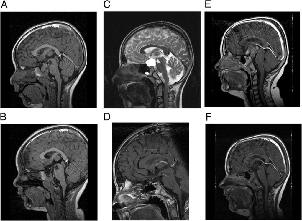
- Imaging tests: To confirm their suspicion, doctors will order imaging tests like a CT scan or an MRI of the brain. These tests provide detailed images of the brain, allowing doctors to see the tumor, its size, and its location.
- Hormone evaluation: Doctors will perform blood tests to check the levels of various hormones produced by the pituitary gland. If the hormone levels are abnormal, it can provide further evidence for the presence of a craniopharyngioma.
- Vision assessment: An eye doctor (ophthalmologist) will check the patient’s vision, including their ability to see details and their peripheral vision. This helps determine if the tumor is affecting the optic nerves, which are responsible for vision.
- Examining the tumor tissue: To make a definitive diagnosis, doctors will need to examine a sample of the tumor tissue under a microscope. This is usually done after a surgical biopsy, where a small piece of the tumor is removed. The tissue examination can confirm the diagnosis and help doctors distinguish between the two main types of craniopharyngioma (adamantinomatous and papillary).
In summary, diagnosing craniopharyngioma involves a combination of examining symptoms, performing imaging tests, checking hormone levels, assessing vision, and analyzing the tumor tissue. Early diagnosis and treatment are important to minimize the risk of long-term complications.
6. Natural History
- Craniopharyngiomas are slow-growing, benign tumors. However, their location near critical brain structures, such as the hypothalamus, pituitary gland, and optic nerves, can cause various health problems if the tumor is not treated. These problems can include headaches, vision loss, hormonal imbalances, and issues with growth and development.
- Over time, the tumor may continue to grow and press on nearby brain structures, causing more severe symptoms and complications. In some cases, the tumor can also block the flow of cerebrospinal fluid, causing a build-up of fluid in the brain (hydrocephalus). This can lead to increased pressure inside the skull, resulting in severe headaches, nausea, vomiting, and cognitive difficulties.
- If left untreated, craniopharyngiomas can cause significant disability and negatively impact a person’s quality of life. In severe cases, the tumor’s growth and related complications can even be life-threatening. That’s why early diagnosis and intervention are critical for managing craniopharyngiomas and minimizing long-term health issues.
7. Treatment Options
Treating craniopharyngiomas typically involves several approaches, depending on the size and location of the tumor, as well as the patient’s overall health. The main treatment options include surgery, radiation therapy, and in some cases, medication. Here’s a simplified explanation of each option:
- Surgery: The primary treatment for craniopharyngiomas is surgical removal of the tumor. The goal is to remove as much of the tumor as possible while minimizing damage to nearby brain structures. There are different surgical approaches, such as endoscopic transnasal surgery (through the nose) or craniotomy (removing a piece of the skull), depending on the tumor’s location and size. Sometimes, complete removal of the tumor is not possible due to its location or risk of damaging vital brain areas.
- Radiation therapy: If the entire tumor cannot be removed during surgery, or if the tumor comes back after surgery, radiation therapy may be used. Radiation therapy involves using high-energy rays to kill tumor cells or stop them from growing. There are several types of radiation therapy, including external beam radiation therapy, stereotactic radiosurgery, and proton therapy, which can be chosen based on the specific case and patient’s needs.
- Medication: In some cases, medication may be used to help manage symptoms or control tumor growth. This can include hormone replacement therapy to address hormonal imbalances caused by the tumor or drugs like interferon or targeted therapies to help slow the growth of the tumor.
- Supportive care: Treatment for craniopharyngiomas can also involve addressing any resulting symptoms or side effects of therapy. This may include physical therapy, occupational therapy, or counselling to help patients cope with the physical and emotional challenges related to their condition.
8. Timing of Surgery
Determining the optimal timing for craniopharyngioma surgery depends on several factors, including the patient’s symptoms, tumor size, location, and overall health. The primary goal of surgery is to remove as much of the tumor as possible while minimizing damage to nearby brain structures. The timing of surgery can impact the success of the procedure and the patient’s long-term outcome.
Early intervention: In many cases, early surgical intervention is recommended, particularly if the patient is experiencing significant symptoms, such as severe headaches, vision loss, or hormonal imbalances. Early surgery can help alleviate these symptoms and prevent further complications caused by the tumor’s growth. Additionally, early surgical intervention may improve the likelihood of complete tumor removal and reduce the risk of recurrence.
Delayed surgery: In some cases, surgery may be delayed if the patient is experiencing only mild symptoms or if the tumor is small and not causing significant problems. This approach is usually taken when the risks associated with surgery are considered to be higher than the potential benefits, such as when the tumor is located in a difficult-to-reach area or near critical brain structures. In such cases, the patient may be closely monitored with regular imaging studies to track the tumor’s growth, and surgery may be performed if symptoms worsen or the tumor grows significantly.
Ultimately, the timing of surgery for craniopharyngioma is determined on a case-by-case basis, considering the patient’s symptoms, the tumor’s characteristics, and the potential risks and benefits of surgery. A multidisciplinary team of specialists, including neurosurgeons, endocrinologists, and radiation oncologists, will work together to determine the most appropriate timing for surgery based on the individual patient’s circumstances.
9. Recovery and Rehabilitation
The recovery and rehabilitation process following craniopharyngioma surgery varies depending on the extent of the tumor, the type of surgery performed, and the individual patient’s overall health. After surgery, patients can expect a period of recovery and rehabilitation to help them regain their strength and return to their normal activities. This process may involve several components:
- Hospital stay: Immediately following surgery, patients typically stay in the hospital for a few days to a week for observation and monitoring. During this time, the medical team will assess the patient’s recovery progress, manage any postoperative pain or complications, and ensure that the patient is stable enough to be discharged.
- Follow-up appointments: After discharge from the hospital, patients will have regular follow-up appointments with their medical team to monitor their recovery, address any lingering symptoms, and check for potential tumor recurrence. These appointments may involve imaging studies, such as MRI or CT scans, and assessments of hormonal levels to ensure that any endocrine imbalances are appropriately managed.
- Physical therapy: Some patients may experience muscle weakness, balance issues, or coordination difficulties following surgery. In these cases, physical therapy can help patients regain their strength and mobility through targeted exercises and activities.
- Occupational therapy: Patients may require assistance with activities of daily living, such as dressing, bathing, or eating, during their recovery. Occupational therapy can help patients regain their independence and adapt to any physical or cognitive limitations resulting from surgery.
- Vision rehabilitation: If the patient has experienced vision loss due to the tumor or surgery, vision rehabilitation specialists can provide strategies and techniques to help patients adapt to their visual impairment and maintain their quality of life.
- Counseling and support: Coping with a craniopharyngioma diagnosis and the challenges of recovery can be emotionally difficult for patients and their families. Counseling and support services can provide emotional support, guidance, and resources to help patients and their loved ones navigate the recovery process.
The duration and intensity of the recovery and rehabilitation process will depend on the patient’s individual needs and the complexity of their surgery. A multidisciplinary team of healthcare professionals, including neurosurgeons, endocrinologists, physical therapists, occupational therapists, and counselors, will collaborate to develop a personalized rehabilitation plan to help the patient achieve the best possible outcome.
10. Outcome
The outcome of craniopharyngioma treatment depends on various factors, such as the tumor’s size, location, extent of surgical resection, and the patient’s overall health. Here are some key aspects to consider when discussing the outcome of craniopharyngioma treatment:
- Surgical success: The primary goal of surgery is to remove as much of the tumor as possible while minimizing damage to nearby brain structures. A successful surgery with complete or near-complete removal of the tumor can improve the patient’s symptoms and reduce the risk of recurrence. However, in some cases, complete removal may not be possible due to the tumor’s location or the risk of damaging vital brain areas.
- Recurrence: Craniopharyngiomas can sometimes recur after initial treatment, especially if complete tumor removal was not achieved. Patients should undergo regular follow-up appointments with their medical team to monitor for potential tumor recurrence, which may require additional treatment, such as radiation therapy or further surgery.
- Long-term complications: Patients who undergo craniopharyngioma surgery may experience long-term complications, such as hormonal imbalances, vision problems, or cognitive difficulties. These complications can result from the tumor itself, damage to nearby brain structures during surgery, or side effects from radiation therapy. Managing these complications may require ongoing medical care, including hormone replacement therapy, vision rehabilitation, or cognitive therapy.
- Quality of life: The overall outcome of craniopharyngioma treatment should be evaluated in terms of the patient’s quality of life. Successful treatment can alleviate symptoms, improve daily functioning, and enhance the patient’s well-being. A multidisciplinary approach, involving various healthcare professionals, is essential for addressing the patient’s physical, emotional, and cognitive needs during their recovery and rehabilitation.
Thusthe outcome of craniopharyngioma treatment depends on multiple factors, including the success of the surgery, the risk of recurrence, and the management of long-term complications. A comprehensive, personalized approach to care, involving a team of specialists, is crucial for achieving the best possible outcome for each patient.
11. Follow-up
Follow-up care is essential for patients who have undergone treatment for craniopharyngioma to monitor their recovery, manage any long-term complications, and detect potential tumor recurrence. Regular follow-up appointments with a multidisciplinary team of healthcare professionals, including neurosurgeons, endocrinologists, and radiation oncologists, will help ensure optimal care and support during the recovery process. Key aspects of follow-up care for craniopharyngioma patients include:
- Imaging studies: Periodic imaging studies, such as MRI or CT scans, are an essential part of follow-up care to monitor the patient’s brain for any signs of tumor recurrence or growth. The frequency of these imaging studies will be determined by the patient’s specific circumstances and the recommendations of their medical team.
- Hormonal evaluation: Regular assessment of pituitary hormone levels is important for patients who have undergone craniopharyngioma treatment, as hormonal imbalances can result from the tumor itself or from surgery or radiation therapy. Appropriate management of hormone levels, which may include hormone replacement therapy, is crucial for maintaining overall health and well-being.
- Visual assessment: Regular ophthalmologic evaluations, including visual acuity and visual field testing, are necessary for patients who have experienced vision problems related to their craniopharyngioma. Ongoing visual assessment helps monitor the patient’s vision and can identify any changes that may require further intervention or rehabilitation.
- Neurological monitoring: Follow-up appointments with a neurologist or neurosurgeon can help monitor the patient’s neurological status, evaluate their recovery progress, and address any ongoing or new symptoms that may arise.
- Rehabilitation and support services: Continued access to rehabilitation and support services, such as physical therapy, occupational therapy, and counseling, is vital for patients recovering from craniopharyngioma treatment. These services help patients regain their strength, independence, and quality of life as they adapt to any physical, cognitive, or emotional challenges resulting from their condition.
In summary, regular follow-up care is essential for patients who have undergone craniopharyngioma treatment to monitor their recovery, manage long-term complications, and detect potential tumor recurrence. A multidisciplinary team of healthcare professionals will work together to provide comprehensive, personalized care throughout the follow-up period to support the patient’s overall health and well-being.
12. Summary
Craniopharyngiomas are rare, benign brain tumors that typically develop near the pituitary gland and hypothalamus, affecting critical brain structures and causing a variety of symptoms, including headaches, vision problems, and hormonal imbalances. The tumors can occur in both children and adults, with different incidence rates and tumor types observed in these age groups.
The exact cause of craniopharyngiomas remains unknown, but they are believed to result from embryonic tissue abnormalities. Diagnosis involves a combination of evaluating the patient’s symptoms, performing imaging tests, assessing hormone levels, examining vision, and analyzing a sample of the tumor tissue.
The natural history of craniopharyngiomas involves slow growth, with progressive symptoms that can lead to severe health problems and reduced quality of life if left untreated. Treatment options for craniopharyngiomas primarily involve surgery to remove the tumor, radiation therapy, and medications to manage symptoms or control tumor growth.
Determining the timing of surgery depends on factors such as the patient’s symptoms, tumor size, location, and overall health. After surgery, patients may require a period of recovery and rehabilitation involving physical therapy, occupational therapy, vision rehabilitation, and counselling.
The outcome of craniopharyngioma treatment is influenced by factors like the success of surgery, the risk of recurrence, and the management of long-term complications. Regular follow-up care is essential for monitoring the patient’s recovery, managing ongoing complications, and detecting potential tumor recurrence.
In conclusion, craniopharyngiomas are complex brain tumors that require a multidisciplinary approach to diagnosis, treatment, and follow-up care. Collaboration among a team of healthcare professionals, including neurosurgeons, endocrinologists, radiation oncologists, and rehabilitation specialists, is crucial to achieving the best possible outcomes for patients with craniopharyngiomas.
13. Disclaimer
This article provides general information about healthcare topics to help individuals make informed decisions and connect with medical professionals for support. However, it is important to note that the information in this article is not a substitute for professional medical advice, diagnosis, or treatment. It is recommended to always seek the advice of a qualified healthcare provider for any medical questions or concerns. Reliance on any information provided in this article is solely at your own risk. If you are interested in scheduling an appointment with a qualified specialist in Pediatric neurosurgery, you can contact us via phone or message on Telegram / WhatsApp at +91 8109 24 7 365.
