1. Introduction
Chiari malformations are a group of structural abnormalities in the base of the skull and cerebellum, the part of the brain responsible for balance and coordination. These malformations cause the cerebellum and, in some cases, the brainstem to descend into the spinal canal, leading to a range of neurological symptoms and complications. There are four types of Chiari malformations, classified based on the severity and specific anatomical abnormalities. Type I is the most common and least severe, while Types II, III, and IV are progressively more severe and less common.
2. Causes
The exact cause of Chiari malformations is not entirely understood. However, they are believed to result from a combination of genetic and environmental factors. The primary cause is thought to be an underdeveloped or abnormally shaped skull, which can be due to genetic mutations, maternal exposure to harmful substances during pregnancy, or other unknown factors. In some cases, Chiari malformations can also be associated with other genetic syndromes or congenital conditions, such as spina bifida.
3. Epidemiology
Chiari malformations are relatively rare, with an estimated prevalence of 1 in every 1,000 to 5,000 individuals, depending on the type. Type I is the most common form, while Types II, III, and IV are much rarer. Chiari malformations can occur in people of any age, but symptoms typically become more noticeable in adolescence or early adulthood for Type I, while Types II, III, and IV often present with symptoms at birth or during early childhood.
4. Symptoms
The symptoms of Chiari malformations vary depending on the type and severity of the condition. Some individuals with Type I Chiari malformation may have no symptoms at all, while others may experience:
- Headaches, often aggravated by coughing, sneezing, or straining
- Neck pain
- Dizziness or balance problems
- Tingling and/or numbness in the hands and feet
- Difficulty swallowing or speaking
- Sleep apnea or abnormal breathing during sleep
In more severe cases, such as Types II, III, and IV Chiari malformations, symptoms can include:
- Severe headache and neck pain
- Breathing difficulties
- Weakness in the arms and legs
- Developmental delays
- Seizures
- Vision problems
5. Pointers to Diagnosis
Diagnosing Chiari malformations typically involves several steps:
- Medical history and physical examination: A healthcare provider will assess the patient’s symptoms, perform a neurological examination, and check for signs of neurological dysfunction.
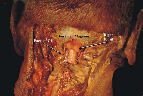
- Imaging studies: Magnetic resonance imaging (MRI) scans are the primary diagnostic tool used to visualize the cerebellum, brainstem, and spinal canal, allowing healthcare providers to identify the presence and severity of the malformation.
6. Natural History
Without appropriate treatment, Chiari malformations can result in a range of complications, including:
- Hydrocephalus: The build-up of cerebrospinal fluid in the brain, leading to increased pressure and potential brain damage
- Spinal cord damage: The compression of the spinal cord can lead to neurological dysfunction and paralysis.
- Syringomyelia: The development of a fluid-filled cyst within the spinal cord, causing pain, weakness, and sensory loss.
- Tethered spinal cord syndrome: A condition where the spinal cord is abnormally attached to the surrounding tissue, leading to neurological symptoms and spinal deformities.
7. Treatment Options
The treatment for Chiari malformations depends on the severity of the condition and the presence of symptoms. Conservative management, including pain management and monitoring, may be appropriate for individualswith mild or asymptomatic cases. In more severe cases or when symptoms significantly impact the individual’s quality of life, surgical intervention may be necessary. There are two main surgical procedures:
- Posterior fossa decompression surgery: This procedure involves removing a small section of the skull and sometimes part of the top vertebrae to create more space for the cerebellum and alleviate pressure on the brainstem and spinal cord. In some cases, the surgeon may also open the protective membrane surrounding the brain (dura mater) to further relieve pressure.
- Spinal cord untethering: In cases where a tethered spinal cord is present, surgery may be performed to release the spinal cord from its abnormal attachments and prevent further neurological damage.
The choice of treatment depends on factors such as the patient’s age, the severity of the malformation, and the individual’s overall health. In some cases, additional treatments may be necessary, including shunt placement for hydrocephalus or ongoing management of related conditions.
8. Timing of Surgery
The timing of surgical intervention for Chiari malformations depends on various factors, such as the severity of symptoms, the presence of complications, and the patient’s overall health. In cases where symptoms are mild or absent, surgery may be delayed and the patient monitored closely for any changes in their condition. In cases where symptoms are severe, progressive, or causing significant disability, surgery may be recommended as soon as possible.
9. Recovery and Rehabilitation
Following surgery, patients typically stay in the hospital for 2-4 days under close observation. During this time, pain medication, antibiotics, and other supportive measures may be administered to aid their recovery. In most cases, intensive care unit (ICU) admission is not necessary, but the patient may require close monitoring by healthcare providers.
Once discharged from the hospital, patients will need to attend regular follow-up appointments with their doctor to monitor their progress. Some patients, particularly those who have experienced neurological damage related to the malformation or its complications, may require rehabilitation to aid in their recovery. This can include physical therapy, occupational therapy, and speech therapy, as needed.
10. Outcome
The outcome for individuals with Chiari malformations depends on various factors, such as the severity of the condition, the timeliness of treatment, and the presence of any complications. With early diagnosis and appropriate surgical intervention, many individuals can experience significant improvement in their symptoms and lead fulfilling lives. However, some individuals may continue to experience neurological deficits or require ongoing medical care and therapy.
11. Follow-up
Long-term follow-up and monitoring are essential for individuals with Chiari malformations. Regular check-ups with healthcare providers, including neurosurgeons and neurologists, help ensure proper recovery and address any potential complications. Additionally, individuals with Chiari malformations may require ongoing evaluations and interventions from other specialists, such as physical therapists, occupational therapists, and speech therapists, to address any lingering neurological issues.
12. Summary
Chiari malformations are a group of structural abnormalities in the base of the skull and cerebellum that can cause a range of neurological symptoms and complications. Early diagnosis, appropriate surgical intervention, and comprehensive long-term care are crucial for improving outcomes and helping individuals with Chiari malformations lead more fulfilling lives. By understanding the causes, symptoms, and treatments of Chiari malformations, individuals and their families can better advocate for their care and manage the challenges associated with this condition.
13. Disclaimer
This website provides general information about healthcare topics to help individuals make informed decisions and connect with medical professionals for support. However, it is important to note that the information on this website is not a substitute for professional medical advice, diagnosis, or treatment. It is recommended to always seek the advice of a qualified healthcare provider forany medical questions or concerns. Reliance on any information provided on this website is solely at your own risk. If you are interested in scheduling an appointment with a qualified specialist in neurosurgery, you can contact us via phone or message on Telegram / WhatsApp at +91 8109 24 7 365.
